
Detecting oral tumors in dogs is a stressful finding for owners. Fortunately, they are not as common as tumors affecting the skin or other body systems. Oral tumors account for 6% of all tumors in the dog. Unfortunately, when they occur, they need to be addressed quickly and appropriately. Approximately 45% of oral tumors in dogs are malignant, with a concern for metastasis to additional body systems. The biggest challenge with oral tumors is simply knowing if one is present. Unless routine (daily to every other day) oral home care is a habit, detection may not be as obvious as a tumor affecting the outside of the body.
Oral tumors are classified as benign or malignant. Oral tumors can be found on the gums, teeth, tongue, and oral mucosa, as well as within the jawbone. An oral tumor is more often seen in older patients.
If an oral mass is detected, the following information should be gathered:
- Biopsy. This is extremely important. Determining the tumor cell type with a biopsy procedure is imperative in planning a curative resolution.
- Obtain full-mouth intraoral dental radiographs or cone-beam CT imaging. Oral imaging is important because a tumor can be more involved in tissues that are not easily viewed. Dental imaging, in the form of dental radiographs, cone-beam CT, conventional CT, or MRI, will be recommended based on the exam findings. Unfortunately, skull radiographs are not as helpful as we would like, and they often do not detect pathology in sufficient detail.
The information obtained from biopsy and imaging will aid in determining whether the oral tumor has the potential for metastasis.
Symptoms associated with oral tumors typically include the following:
- Facial swelling
- Oral malodor
- Mobile teeth
- Decreased appetite
- Oral bleeding
- Oral pain
- Weight loss
- Non-healing recent dental extraction site
- Enlarged mandibular lymph nodes
It is important to note that the above clinical signs may also be seen with periodontal disease. Oral pathology signs quite frequently overlap, necessitating the importance of an exam with a veterinarian or a board-certified veterinary dental specialist.
Types of Oral Tumors in Dogs
Oral tumors in dogs are divided into two categories: benign and malignant. Benign tumors may be inflammatory or odontogenic in nature. Odontogenic refers to arising from tooth structure.
1. Benign Oral Tumors
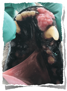
- Gingival hyperplasia. Gingival hyperplasia manifests as gingival overgrowth and is composed of fibrous tissue. It is a common occurrence in shorter-faced dogs (brachycephalic breeds) such as bulldogs, pugs, and boxers.
Trivia! Gingival hyperplasia occurs in humans who tend to breathe with their mouths open. The chronic exposure of air to the oral cavity predisposes the oral tissues to become thickened and overgrown as a protective barrier. In dogs with smaller muzzles, a shorter nasal passage will trigger breathing through the mouth as a compensatory measure.
Medications can cause gingival hyperplasia. Certain medications, such as cyclosporine and amlodipine are known causes of gingival hyperplasia. Treatment involves the surgical removal of the excess tissue, which is called a gingivectomy. Surgical treatment is followed up by routine tooth brushing to minimize reoccurrence.
Gingival hyperplasia covering the maxillary incisors
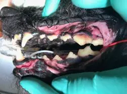
- Focal fibrous hyperplasia. Reactive gum tissue may occur in response to chronic periodontal disease and may affect single or multiple teeth. Routine dental care and oral home care are beneficial in preventing recurrence.
Fibrous hyperplasia in response to plaque and tartar at the gingival margin
- Peripheral Odontogenic Fibromas (POF): These tumors originate from the tissues around the teeth and usually affect one tooth. They are slow-growing, and surgical excision is curative. In some cases, the tooth associated with the mass tends to be removed to avoid recurrence.
Peripheral odontogenic fibroma (POF) appearance on a maxillary premolar
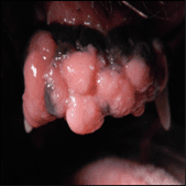
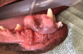
- Papillomas: These are wart-like growths caused by a virus (canine papillomavirus) and are more common in young dogs. They often resolve on their own but may require treatment if they grow large or cause discomfort. They may appear if the immune system is taxed with any concurrent health issues. Common sites of occurrence are the tongue and oral mucosa.
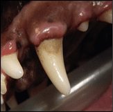
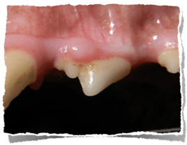
- Canine acanthomatous ameloblastoma (CAA): This is a slow-growing, expansile tumor that occurs most commonly in medium to large breed dogs. Canine acanthomatous ameloblastoma is not considered malignant, BUT it is locally invasive and includes gingiva, oral mucosa, and bone. Surgical removal involves the removal of ALL the tumor, including where it is detected in the jawbone. Canine acanthomatous ameloblastoma is often found at the front of the mouth, affecting the incisor or canine teeth.
CAA causing tooth displacement of the mandibular incisors
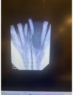
CAA appearance affecting the lower jaw (mandible)
2. Malignant Oral Tumors
- Malignant Melanoma. Malignant melanoma is the most common malignant oral tumor seen in dogs. It may present pigmented (melanotic) or non-pigmented (amelanotic). It is seen in all breeds but may occur more frequently in cocker spaniels. The site of occurrence is anywhere in the oral cavity, but is more often noted in the oral mucosa near the back of the mouth. Malignant melanoma commonly spreads to the lungs and regional lymph nodes. Treatment often involves multiple modalities, including surgical excision, radiation, and follow-up chemotherapy. Consultation with an oncologist, in addition to an oral surgeon, is recommended.

Amelanotic melanoma
Melanotic melanoma
- Squamous Cell Carcinoma (SCC): This is the second most common malignant oral tumor seen in dogs. It is often ulcerative and proliferative in appearance. It often will rapidly invade bone. Oral bleeding is a common symptom, and it is commonly mistaken for periodontal disease. Treatment involves surgical excision and radiation therapy, among other emerging treatment modalities. Consultation with an oncologist, in addition to an oral surgeon, is recommended. *A papillary variant may occur, which is not considered malignant, and surgical excision is expected to be curative. An oncologist consultation for this variant is typically not needed.
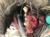
SCC affecting the maxilla (upper jaw) in a small-breed dog
- Fibrosarcoma (FSA): Fibrosarcoma is the third most common malignant oral tumor seen in the canine oral cavity. It is seen more frequently in larger breed dogs such as retrievers. It often presents as a raised, broad-based mass and involves bone as well as oral mucosa. Treatment involves surgical excision with radiation therapy. Biopsy results can be unrewarding, and a repeat biopsy may be needed with cone beam CT or conventional CT imaging for detection. Reoccurrence may occur despite surgical excision. Consultation with an oncologist, in addition to an oral surgeon, is recommended.
FSA affecting the right mandible in a Labrador retriever
Conclusion
Oral masses can be challenging to detect and identify. An anesthetized exam with full mouth dental imaging and a biopsy is recommended if an oral mass is present or suspected. Due to the limited amount of tissue in the mouth and frequent involvement of select tumors in the surrounding bone and tissue, a biopsy is recommended before a more significant surgery. At Montana Pet Dentistry and Oral Surgery, a healthy and comfortable oral cavity is our passion. You can reach us at (406) 599-4789 to schedule a consultation and discuss your canine companion’s oral health concerns.
Images used under creative commons license – commercial use (11/22/2024) Image by congerdesign from Pixabay