
Dental imaging is an essential component of dental care. Dogs have 42 teeth, while cats have 30 teeth. A large percentage of the tooth structure (60%) is below the gumline, and diagnosis of dental disease and oral pathology requires imaging. Many patients have painful problems hidden under the gumline that can only be identified with dental imaging, including advanced options like cone beam CT for pets.


Types of Dental Imaging
The two common forms of dental imaging include dental radiographs and cone beam CT (CBCT) imaging. Skull radiographs have been used in the past, but unfortunately, do not provide the level of detail required to evaluate the teeth and periodontal structures and lack the spatial orientation to differentiate and interpret maxillofacial structures accurately. Limited or select individual dental radiographs during a cleaning procedure often miss oral pathology and can leave untreated problems in the mouth.
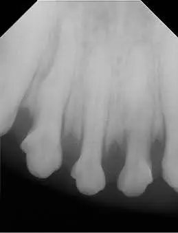
Tooth resorption on dental X-ray

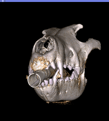
Tooth resorption on CBCT image with a more detailed angle of evaluation
Intraoral Dental Radiographs
Traditionally, dental imaging is achieved with intraoral dental radiographs. Digital dental radiographs have been an integral mainstay in veterinary dentistry, but they do have limitations. A dental radiograph, or X-ray, is a two-dimensional image that provides challenges in evaluating a three-dimensional structure.
Advantages of Cone Beam CT for Pets
Cone beam CT imaging has emerged as a diagnostic improvement to dental radiographs. Obtaining a CBCT scan on our patients takes less time than taking intraoral dental radiographs, which reduces the duration of anesthesia for your pet. Dental pathology may take months to appear on a dental radiograph, whereas it can be detected in as little as a week with cone beam imaging. This allows us to assess areas hidden behind teeth, as well as view an area of interest from different angles, which greatly improves the diagnostic information available when making treatment recommendations.
The three-dimensional image provided by the CBCT scanner helps us visualize any oral pathology that we may not have been able to detect in an X-ray or routine oral exam. This enables us to eliminate doubt when making our diagnosis and coming up with a treatment plan, making cone beam CT for pets a valuable tool in veterinary dentistry.
Cone Beam CT for Advanced Oral Conditions
In cases of maxillofacial trauma, oral tumors, and facial swellings, CBCT imaging can rapidly define the extent of pathology. This, in addition to the ability to produce three-dimensional reconstructed images, makes surgical planning more specific and efficient. In cases of non-vital (dead) teeth, the ability to predict the prognosis of endodontic treatment is improved significantly with the use of cone beam CT imaging.
Comprehensive Dental Health Assessments
In addition to the three-dimensional view, the VetCat Cone Beam CT scanner provides excellent detail and clarity of images. It can be used for finding undetected pathology in seemingly routine procedures such as oronasal fistulas, facial trauma, dental injuries, embedded teeth, areas of bone loss hidden under the gumline, displaced and/or retained roots, nasal tumors, oral masses, TMJ abnormalities, and many other hidden painful problems.
Using the cone beam CT for pets, along with dental X-rays, gives us the most comprehensive view of your pet’s oral health. Montana Pet Dentistry and Oral Surgery is proud to offer cone beam CT imaging as part of all our dental cleaning packages.
Images used under creative commons license – commercial use (07/02/2025) Photo by Krista Mangulsone on Unsplash