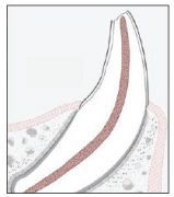
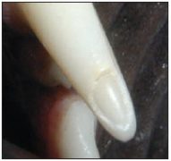
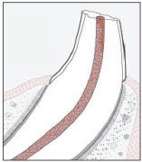
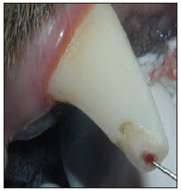
Several different types of tooth fractures may occur in pets, with crown fractures being the most common. There are basically 2 types of crown fractures; Complicated Crown Fractures (charted as CCF) and Uncomplicated Crown Fractures (charted as UCF). Regardless of how the patient is acting or how long the owner thinks the fracture has been present, all fractured teeth should be treated. Your patients will rarely show any sign of pain. When you treat the teeth, however, they almost always show signs of improvement. Selection of the appropriate treatment modalities depends on the type of fracture, radiographic appearance of the tooth and, in some cases, the duration of the fracture.
Complicated Crown Fractures (Pulp Exposure)
Complicated Crown Fractures are fractures in which the pulp (nerve) chamber has become exposed. These teeth should be treated with root canal treatment or extraction. Despite rumors to the contrary, pulp exposure will never spontaneously heal, except in experimental germ-free rats. The presence of bacteria in the oral cavity always results in pulp necrosis and endodontic infection. The brown spots seen on some teeth are stained dentin, and do not represent prior pulp exposure that has healed.
Occasionally, Vital Pulp Therapy (aka- pulp cap therapy) may be used to treat a very recent pulp exposure, but root canal therapy is almost always a better choice and is associated with a much higher success rate. I would only choose Vital pulp therapy over root canal therapy in recent fractures when the patient is 14 months of age or younger.
In these patients, the walls of the root are still very thin, and maintaining the vitality of the tooth allows for continued development and strengthening of the tooth. After 14 months of age, the tooth should be strong enough to last for the life of the patient, and root canal therapy is a more appropriate choice.
Uncomplicated Crown Fractures
In Uncomplicated Cown Fractures, the pulp is not exposed. Because the enamel of dogs and cats is so thin, these fractures almost always result in exposure of the dentin, which makes up around 80% of the tooth structure.
Dentin appears to be a solid substrate, but it contains 40,000 plasma filled tubules/mm2. Exposure of these dentin tubules is painful, and the tubules are large enough to allow direct bacterial migration into the pulp chamber, resulting in endodontic infection. What appears to be a solid substrate is not.
These teeth should all be radiographed and assessed for vitality. If they are still vital (alive) they may be treated by smoothing the fracture site and applying bonded resin sealants to the site. The bonded sealants block the dentin tubules, decrease pain, help prevent bacterial migration into the pulp chamber, and assist the tooth in healing itself from inside the pulp chamber. We teach this technique in our Level II class.
Do’s & Don’ts of Treating Dog Tooth Fractures
Bonded sealants should not be performed in complicated crown fractures or teeth with near exposure of the pulp, as you will trap bacteria inside the tooth, leading to endodontic infection and death of the tooth. If a tooth with an uncomplicated crown fracture is found to be non-vital or infected inside, then it should be treated with r
oot canal therapy or extraction. You can never tell what treatment is appropriate unless you utilize dental radiographs. In many cases, restoration of normal anatomy with composite restorative materials is also indicated, which can serve to restore full function and strength.
The amount of visible damage to the tooth is often a poor predictor of whether or not the pulp has been compromised. Some large fractures may result in no pulp pathology and some small fractures result in pulp necrosis that lead to an abscess. Fractured and worn teeth require careful clinical and radiographic evaluation to determine what treatment, if any, is indicated. Bonded sealants or composite restoration of a tooth does not ensure that the tooth will not abscess! Follow-up radiographs should be obtained in 6-12 months and periodically thereafter to verify that treated teeth remain healthy.
Do:
- Radiograph all fractured, discolored or worn teeth.
- Offer root canal treatment as an alternative to extraction for complicated crown fractures or uncomplicated fractures in which the tooth has become non-vital or endodontically infected.
- Chart and note all tooth injuries on the patient’s dental chart.
- Recheck all treated teeth regularly with dental radiographs.
Don’t:
- Assume if tooth fracture “looks minor” that no injury or infection of the pulp has occurred. Always obtain dental radiographs.
- Perform bonded sealants or restorative treatment without obtaining quality dental radiographs first.
- Perform bonded sealants or other restorative treatment without advising recheck dental radiographs in 6 months.
- Perform bonded sealants on teeth with exposed pulp (complicated crown fractures).
Take-Home Message: Improper diagnostics and dental restorative treatment may doom the pet to continued pain and infection because symptoms of dental pain in dogs and cats are not noted in most cases. The radiographic signs of teeth that are non-vital or endodontically infected can be very subtle or non-existent, taking years to develop in some cases. Abscessed teeth rarely swell up or have any associated drainage.
A dog with an improperly treated fractured limb will continue to limp and show obvious signs of the ineffective treatment. A dog with an improperly treated fractured tooth that is non-vital or has abscessed rarely show signs of pain. They continue to eat, drink and play, but live with chronic subclinical pain and infection. This will be illustrated dramatically in next month’s case report.
Download Printable Newsletter (PDF)
Photo by Aditya Joshi on Unsplash (2/12/2021)
